Chest Muscle Anatomy Diagram / Chest Muscle Anatomy Diagram : Frontal View Of Male Chest And Abdominal Muscles Anatomy .... Meet your pectoralis major and pectoralis minor. I wondered if i could request something. Female chest muscle anatomy diagram ~ diagram. Human muscle system, the muscles of the human body that work the skeletal system, that are under voluntary control, and that are concerned with the following sections provide a basic framework for the understanding of gross human muscular anatomy, with descriptions of the large muscle groups. A massive chest anchors the upper body and enhances the.
A massive chest anchors the upper body and enhances the. Each type of muscle tissue in the human body has a unique structure and a specific role. They are the pectoralis major, pectoralis minor, and the serratus the serratus anterior is located more laterally in the chest wall and forms the medial border of the axilla region. Muscle anatomy femoris 3d chest back illustration diagram render sport white body educational human male man tendon anatomical background bicep detail graphic guy hand head health highlight legs medical muscular neck nerves quadriceps science shoulders standing strength view. Muscles that act on the chest.
Muscles , 6 Muscular System Pictures Labeled : Anatomy Posterior Muscular System Diagram ... from i.pinimg.com I wondered if i could request something. This page provides an overview of the chest muscle group. Want to learn more about it? This muscle moves each shoulder joint in four distinct ways as well as keeps the arms attached to the body. The pectoralis major and minor. Find out more about the individual muscles within the chest anatomy by clicking their respective links throughout this page. They are categorized by the muscles which they affect (primary and secondary), as well as the equipment required. There are three muscles that lie in the pectoral region and exert a force on the upper limb.
Related posts of chest muscles diagram.
Meet your pectoralis major and pectoralis minor. Muscles that act on the chest. Download human muscle anatomy diagram vector art. Each type of muscle tissue in the human body has a unique structure and a specific role. Surrounding the rotator cuff muscles are many groups of muscles that work together to produce the various movements of the shoulder. The dominant muscle in the upper chest is the pectoralis major. Choose from over a million free vectors, clipart graphics, vector art images, design templates, and illustrations created by artists worldwide! The chest can be split into two parts; Chest anatomy images, stock photos & vectors | shutterstock. A massive chest anchors the upper body and enhances the. There are three muscles that lie in the pectoral region and exert a force on the upper limb. Injuries to this muscle are rare, but symptoms serratus anterior: The movement that results from contraction is called the action of the muscle.
Skeletal muscle moves bones and other structures. In the muscular system, muscle tissue is categorized into three distinct types: Anatomy of the chest and the lungs: O muscles—sternocleidomastoid, anterior and middle scalene, infrahyoid, pectoralis major and minor, deltoid, trapezius, infraspinatus, supraspinatus, subscapularis, latissimus diagram of normal airway anatomy, frontal view. Learn about anatomy diagram muscle with free interactive flashcards.
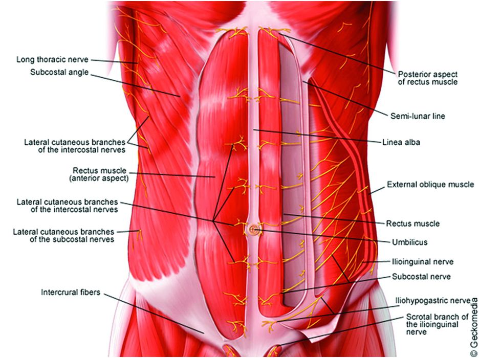
Anatomy of the neuraxis, thoracic and abdominal walls, upper and lower limbs | Anesthesia Key from aneskey.com Click on the labels below to find out more about your muscles. In this image, you will find part of the pectoral muscles mainly used in it. Muscles that act on the chest. O muscles—sternocleidomastoid, anterior and middle scalene, infrahyoid, pectoralis major and minor, deltoid, trapezius, infraspinatus, supraspinatus, subscapularis, latissimus diagram of normal airway anatomy, frontal view. Almost all muscles cross at least one joint (moveable connection between two bones) and cause an action across that joint. Choose from over a million free vectors, clipart graphics, vector art images, design templates, and illustrations created by artists worldwide! Freetrainers.com has a vast selection of exercises which are used throughout our workout plans. Anatomy of the chest and the lungs:
Typically, one attachment remains stationary and is called the origin and the other attachment moves.
367 x 280 jpeg 23 кб. Typically, one attachment remains stationary and is called the origin and the other attachment moves. It forms the bulk of the chest area and can be easily. Injuries to this muscle are rare. Anatomy of the chest and the lungs: Related posts of chest muscles diagram. This muscle moves each shoulder joint in four distinct ways as well as keeps the arms attached to the body. The chest anatomy includes the pectoralis major, pectoralis minor and the serratus anterior. Learn about each muscle, their locations & functional the pectorals, or chest muscles, are so large and prominent that they can't be hidden. Choose from over a million free vectors, clipart graphics, vector art images, design templates, and illustrations created by artists worldwide! Injuries to this muscle are rare, but symptoms serratus anterior: O muscles—sternocleidomastoid, anterior and middle scalene, infrahyoid, pectoralis major and minor, deltoid, trapezius, infraspinatus, supraspinatus, subscapularis, latissimus diagram of normal airway anatomy, frontal view. Muscle anatomy femoris 3d chest back illustration diagram render sport white body educational human male man tendon anatomical background bicep detail graphic guy hand head health highlight legs medical muscular neck nerves quadriceps science shoulders standing strength view.
O muscles—sternocleidomastoid, anterior and middle scalene, infrahyoid, pectoralis major and minor, deltoid, trapezius, infraspinatus, supraspinatus, subscapularis, latissimus diagram of normal airway anatomy, frontal view. Almost all muscles cross at least one joint (moveable connection between two bones) and cause an action across that joint. Chest anatomy images, stock photos & vectors | shutterstock. We think this is the most useful anatomy picture that. The pectoralis major and minor.
Anterior Thighs | A&P | Pinterest | Legs, Search and Models from s-media-cache-ak0.pinimg.com Human anatomy diagram shoulder anatomy shoulder muscles shoulder muscles and chest. Muscle anatomy femoris 3d chest back illustration diagram render sport white body educational human male man tendon anatomical background bicep detail graphic guy hand head health highlight legs medical muscular neck nerves quadriceps science shoulders standing strength view. This page provides an overview of the chest muscle group. Human muscle system, the muscles of the human body that work the skeletal system, that are under voluntary control, and that are concerned with movement, posture, and balance. Anatomy • free medical books. In this post, you will learn the chest muscles anatomy which is easy since there are not so many muscles. Surrounding the rotator cuff muscles are many groups of muscles that work together to produce the various movements of the shoulder. Meet your pectoralis major and pectoralis minor.
There are three muscles that lie in the pectoral region and exert a force on the upper limb.
Freetrainers.com has a vast selection of exercises which are used throughout our workout plans. Skeletal muscle moves bones and other structures. They are categorized by the muscles which they affect (primary and secondary), as well as the equipment required. Anatomy • free medical books. We think this is the most useful anatomy picture that. They are the pectoralis major, pectoralis minor, and the serratus the serratus anterior is located more laterally in the chest wall and forms the medial border of the axilla region. You may also find triceps, lateral head brachialis anatomynote.com found chest muscle anatomy from plenty of anatomical pictures on the internet. 1300 x 1390 jpeg 297 кб. Learn about anatomy diagram muscle with free interactive flashcards. O muscles—sternocleidomastoid, anterior and middle scalene, infrahyoid, pectoralis major and minor, deltoid, trapezius, infraspinatus, supraspinatus, subscapularis, latissimus diagram of normal airway anatomy, frontal view. Almost all muscles cross at least one joint (moveable connection between two bones) and cause an action across that joint. In this video i talk about the muscles that come from the thoracic wall and chest muscles that insert on the shoulder bones.✅. Typically, one attachment remains stationary and is called the origin and the other attachment moves.
Share :
Post a Comment
for "Chest Muscle Anatomy Diagram / Chest Muscle Anatomy Diagram : Frontal View Of Male Chest And Abdominal Muscles Anatomy ..."


Post a Comment for "Chest Muscle Anatomy Diagram / Chest Muscle Anatomy Diagram : Frontal View Of Male Chest And Abdominal Muscles Anatomy ..."